
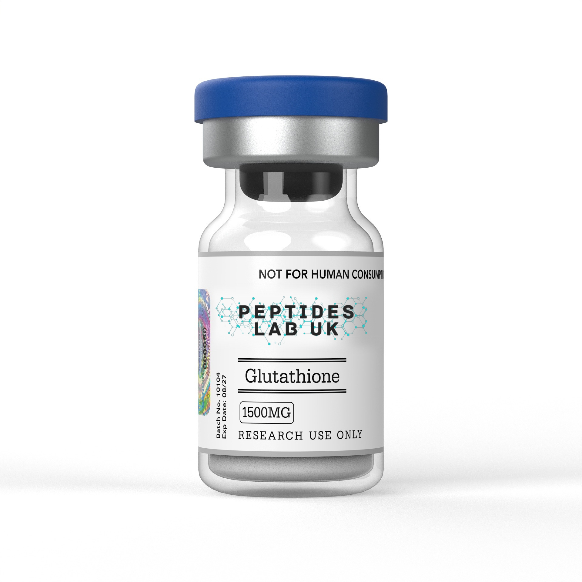
Glutathione (gamma-L-glutamyl-L-cysteinylglycine, GSH) is an endogenous tripeptide composed of glutamate, cysteine and glycine, and it is the most abundant low-molecular-weight thiol antioxidant in mammalian cells. Its biological activity centres on the reactive sulfhydryl (-SH) group contributed by its cysteine residue, which allows glutathione to act as an electron donor in redox reactions and a nucleophile in xenobiotic conjugation. The ratio of reduced glutathione (GSH) to its oxidised disulfide form (GSSG) is a widely used indicator of cellular redox status in laboratory research, with normal GSH/GSSG ratios reported at approximately 100:1 in the cytoplasm, though considerably lower in other cellular compartments. Within laboratory research, glutathione is used extensively as a tool compound for studying oxidative stress, cellular detoxification pathways and mitochondrial redox biology.
What Is Glutathione?
Glutathione is a tripeptide with the molecular formula C10H17N3O6S and a molecular weight of approximately 307 Da, consisting of L-glutamate, L-cysteine and glycine linked through an unusual peptide architecture. Unlike conventional peptides, in which amino acids are joined through standard alpha-peptide bonds, glutathione’s glutamate residue is linked to cysteine through a gamma-peptide bond, connecting the gamma-carboxyl group of glutamate rather than its alpha-carboxyl group. This structural feature is functionally significant: the gamma-peptide bond protects glutathione from cleavage by most intracellular peptidases, which recognise and cleave conventional alpha-peptide bonds, contributing substantially to glutathione’s metabolic stability relative to standard tripeptides.
Glutathione biosynthesis proceeds through a two-enzyme pathway. The first and rate-limiting step is catalysed by glutamate-cysteine ligase (GCL), which joins glutamate and cysteine through the gamma-peptide bond to form gamma-glutamylcysteine. The second step is catalysed by glutathione synthetase, which adds glycine to complete the tripeptide structure. This biosynthetic pathway is tightly regulated in response to cellular redox status, and cysteine availability is generally considered the rate-limiting substrate for glutathione synthesis under most physiological conditions, since glutamate and glycine are typically present in comparative excess.
Glutathione is compartmentalised across multiple cellular locations, with distinct redox environments maintained in each. Research has reported that the GSH/GSSG ratio, a key indicator of local redox status, differs substantially by compartment: approximately 100:1 in the cytoplasm, approximately 10:1 in mitochondria, and considerably lower, in the range of 1:1 to 3:1, in the endoplasmic reticulum, reflecting the more oxidising environment required for correct disulfide bond formation during protein folding in that compartment. This compartmentalisation is directly relevant to laboratory research design, since glutathione’s functional role and redox behaviour can differ meaningfully depending on which subcellular compartment is under investigation.
The distinction between reduced GSH and oxidised GSSG is central to understanding glutathione’s research applications. GSH is the biologically active, reduced monomeric form, in which the cysteine thiol group is available to donate electrons or participate in nucleophilic reactions. GSSG is the oxidised disulfide dimer, formed when two GSH molecules are joined through a disulfide bond following electron donation, typically during neutralisation of reactive oxygen species. Under conditions of significant oxidative stress, the cellular capacity to reduce GSSG back to GSH, a process dependent on glutathione reductase and NADPH, can become overwhelmed, leading to GSSG accumulation and a measurable shift in the GSH/GSSG ratio, which researchers use as a quantitative marker of oxidative stress severity in experimental systems.
Mechanism of Action
The principal mechanism attributed to glutathione is direct and enzymatically catalysed neutralisation of reactive oxygen species (ROS). Aerobic cellular metabolism generates hydrogen peroxide as a byproduct, and this is metabolised by glutathione peroxidase (GPx) in the cytosol and mitochondria, using GSH as the electron donor and generating GSSG as the oxidised byproduct. This GPx-catalysed reaction is one of the principal cellular routes for hydrogen peroxide detoxification, operating alongside catalase-mediated peroxide breakdown in the peroxisome. Organic peroxides can similarly be reduced using either glutathione peroxidase or glutathione S-transferase, providing a degree of enzymatic redundancy in the cellular peroxide-detoxification system.
Nucleophilic conjugation via glutathione S-transferases (GSTs) represents a second major mechanistic pathway, distinct from direct ROS neutralisation. GSTs catalyse the conjugation of GSH to a wide range of electrophilic xenobiotic compounds, including certain drug metabolites, environmental toxins and reactive intermediates generated during Phase I metabolism, forming glutathione conjugates that are generally less reactive and more readily exported from the cell than their unconjugated precursors. This GST-mediated conjugation pathway is central to cellular xenobiotic detoxification and has made glutathione and its associated enzyme family a standard tool in toxicology and pharmacology research examining Phase II drug metabolism.
Mitochondrial membrane protection represents a further mechanistic dimension of glutathione’s research profile. Because mitochondria are a major site of ROS generation, arising as a byproduct of electron transport chain activity, mitochondrial GSH pools play a particularly important role in limiting oxidative damage to mitochondrial membrane lipids, proteins and DNA. Research has reported that mitochondrial GSH is maintained through active transport from the cytosol rather than local synthesis, since the enzymes required for glutathione biosynthesis are not present within mitochondria themselves, making mitochondrial GSH transport a distinct area of research interest relative to cytosolic glutathione metabolism.
Glutathione also participates in a broader antioxidant recycling network, contributing to regeneration of other cellular antioxidants, including ascorbic acid (vitamin C) and, indirectly, vitamin E, through interconnected redox cycling reactions. This positions glutathione not merely as an independent antioxidant but as a central hub within a coordinated cellular antioxidant defence network, a property relevant to researchers studying combined or comparative antioxidant depletion and repletion protocols.
Redox-sensitive transcription factor modulation represents a further mechanistic layer increasingly documented in the literature. Beyond its direct antioxidant and conjugation functions, glutathione participates in protein redox signalling through reversible post-translational modifications, including S-glutathionylation, in which GSH forms a mixed disulfide with protein cysteine residues, protecting them from irreversible oxidative damage while also modulating protein function and downstream signalling. This glutathionylation-dependent regulation has been documented to intersect with redox-sensitive transcription factor activity, connecting glutathione’s biochemistry to broader gene-expression regulation beyond its role as a stoichiometric antioxidant.
What the Research Shows
A foundational review of glutathione biosynthesis has consolidated the biochemical pathway through which GCL and glutathione synthetase produce GSH, describing the GPx- and catalase-mediated routes for hydrogen peroxide detoxification, the GSSG reductase-mediated regeneration cycle dependent on NADPH, and the mechanisms by which cells manage GSSG accumulation under conditions where reduction capacity is overwhelmed, including active GSSG export and mixed disulfide formation with protein thiols (glutathione synthesis and redox cycle review).
A separate review examining glutathione’s role in redox signalling has detailed the biosynthetic pathway in greater mechanistic depth, describing S-glutathionylation as the conjugation of GSH with protein thiol residues and summarising evidence for glutathione’s broader role in cell signalling beyond its classical antioxidant function, including its interaction with peroxiredoxins, which may function as redox sensors within this signalling network (glutathione synthesis and redox signalling review).
A more recent and comprehensive review focused specifically on glutathione’s role in protein redox modulation through S-glutathionylation and S-nitrosylation has characterised glutathione’s gamma-peptide bond structure and its resistance to peptidase cleavage, documented compartment-specific GSH/GSSG ratios across the cytoplasm, mitochondria and endoplasmic reticulum, and examined the interplay between glutathione redox status and thioredoxin-family enzymes, including thioredoxin, glutaredoxin and protein disulfide isomerase, in regulating cell viability through these reversible thiol modifications (glutathione protein redox modulation review).
Hepatocyte injury research has long used glutathione depletion and repletion as a standard experimental paradigm for studying oxidative and xenobiotic-induced liver cell injury, given the liver’s central role in Phase II xenobiotic metabolism and its correspondingly high glutathione turnover, with cultured hepatocyte models frequently used to characterise dose-dependent relationships between glutathione status and cellular susceptibility to oxidative or chemical insult. Neuroprotective pathway research has similarly examined glutathione status in neuronal and glial cell culture models, reflecting the central nervous system’s high metabolic rate and correspondingly elevated vulnerability to oxidative stress, with glutathione depletion protocols used as an experimental tool to model oxidative neuronal injury in preclinical research settings.
Mitochondrial oxidative balance research has documented the distinct regulation of mitochondrial GSH pools relative to cytosolic glutathione, given the absence of glutathione biosynthetic machinery within mitochondria themselves and the resulting dependency on active transport, a research area of particular relevance to studies examining mitochondrial dysfunction and its relationship to broader cellular oxidative stress.
Research Applications
Within laboratory settings, glutathione is used across several well-established redox biology and toxicology research contexts. ROS inhibition assays in cultured cells represent a core application, in which researchers manipulate intracellular GSH levels, either through direct supplementation or through pharmacological depletion using agents such as buthionine sulfoximine, to characterise the relationship between cellular glutathione status and susceptibility to oxidative stressors, using fluorescent or luminescent ROS-detection probes to quantify oxidative burden under controlled experimental conditions.
Xenobiotic detoxification profiling constitutes a further major research application, using hepatocyte or other metabolically active cell models to characterise GST-mediated conjugation of test compounds, an approach standard in pharmacology and toxicology research for assessing Phase II drug metabolism and predicting potential toxicity of reactive drug metabolites. Mitochondrial bioenergetics assays are used to examine the relationship between mitochondrial GSH status and respiratory function, often in combination with oxygen consumption measurement techniques, to characterise how oxidative stress and glutathione depletion affect mitochondrial membrane integrity and electron transport chain efficiency.
Redox kinetics studies represent a further significant application area, in which researchers directly quantify the GSH/GSSG ratio using techniques such as HPLC with electrochemical or fluorescence detection, or enzymatic recycling assays, to track dynamic changes in cellular redox status over time or in response to experimental interventions. When selecting a certified Glutathione research peptide for cellular ROS inhibition or detoxification pathway protocols, researchers should confirm the exact tripeptide structure and purity documentation supplied, since the ratio of reduced to oxidised glutathione in a given batch is directly relevant to reproducing redox-sensitive assay results, and pre-oxidised material can introduce a confounding baseline shift into GSH/GSSG ratio measurements.
Comparative pharmacology work has also examined glutathione alongside other low-molecular-weight thiol antioxidants and related tripeptide compounds, providing researchers with a broader comparative framework for studying structure-activity relationships within cellular thiol-based redox biology.
Purity, Analytical Verification, Storage and Handling
Research-grade glutathione should be accompanied by a certificate of analysis confirming purity by HPLC, typically at or above 98 percent, together with mass spectrometry verification confirming the correct tripeptide structure and the ratio of reduced (GSH) to oxidised (GSSG) forms present in the supplied material. Because glutathione’s reactive thiol group is readily susceptible to spontaneous oxidation on exposure to atmospheric oxygen, analytical documentation specifying the GSH/GSSG composition of a given batch is particularly important for redox-sensitive research applications, where an unexpectedly high baseline GSSG content could confound experimental interpretation. When sourcing high-purity glutathione for redox stability and oxidative stress assays, UK research laboratories must confirm that each batch is backed by this documentation rather than relying on a generic product listing.
Thiol-group oxidation prevention is a central handling consideration throughout the working life of a glutathione batch. Lyophilised glutathione should be stored at -20°C under inert atmosphere where possible, protected from light, moisture and oxygen exposure, since the free thiol group remains susceptible to gradual oxidation even in the solid lyophilised state over extended storage periods. Reconstitution should be carried out using deoxygenated buffers wherever the experimental protocol allows, since dissolved oxygen in aqueous buffer systems can drive spontaneous GSH-to-GSSG conversion during and immediately following reconstitution, particularly at neutral to alkaline pH, where thiol reactivity is enhanced.
Once reconstituted, glutathione solutions should be used promptly and kept refrigerated at 2-8°C if short-term storage is unavoidable, with researchers following supplier-specific guidance regarding the maximum acceptable storage duration before oxidation compromises the GSH/GSSG ratio beyond acceptable limits for a given assay. Aliquoting reconstituted material into single-use volumes under inert gas where practical is recommended to minimise repeated oxygen exposure across an experimental run, since each freeze-thaw and re-opening cycle introduces further opportunity for thiol oxidation.
Frequently Asked Questions
Why is the GSH/GSSG ratio used as a marker of cellular redox status in research?
GSH is the reduced, biologically active form of glutathione, while GSSG is the oxidised disulfide formed after GSH donates electrons to neutralise reactive oxygen species. Because this ratio shifts predictably under oxidative stress, researchers use it as a quantitative indicator of cellular redox balance across cytoplasmic, mitochondrial and endoplasmic reticulum compartments, which each maintain distinct baseline ratios.
How can researchers prevent unwanted glutathione oxidation during assay preparation?
Because glutathione’s reactive thiol group is prone to spontaneous oxidation on exposure to atmospheric oxygen, researchers should use deoxygenated buffers for reconstitution where possible, minimise exposure to neutral-to-alkaline pH conditions that enhance thiol reactivity, and use freshly reconstituted material promptly rather than storing working solutions for extended periods.
What distinguishes glutathione’s peptide bond structure from conventional peptides?
Glutathione’s glutamate residue is joined to cysteine through a gamma-peptide bond rather than the standard alpha-peptide bond found in most peptides. This structural feature protects glutathione from cleavage by conventional intracellular peptidases, contributing to its metabolic stability relative to typical tripeptide sequences.
How should research-grade glutathione be verified before use in a redox assay?
Researchers should request a batch-specific certificate of analysis confirming HPLC purity of 98 percent or higher and mass spectrometry confirmation of the correct tripeptide structure, along with documentation of the GSH/GSSG ratio present in the batch, since baseline oxidation status is directly relevant to reproducing redox-sensitive experimental findings.
Glutathione, as supplied by Peptides Lab UK and comparable UK research suppliers, is intended strictly for in-vitro and animal-model laboratory research. It is not licensed or intended for human or veterinary use, and nothing in this article should be interpreted as guidance for personal administration.